Sunday 28 February 2016

B Sc III 

Genetic Engineering

follow links to know more

http://agbiosafety.unl.edu/basic_genetics.shtml

http://pamelaronald.blogspot.in/2008/08/basic-concepts-in-science-crop-genetic.html

http://www.encyclopedia.com/topic/genetic_engineering.aspx
 

Saturday 13 February 2016

B Sc II Gnetales - Gnetum


Gnetum plant
 
Gnetum leaf resembles angosperms
 
Gnetum strobilus (male flowers)
 
Gnetum strobilus (female flowers)
 
Gnetum seeds


Saturday 6 February 2016

B Sc I , B Sc II  and B Sc III Practical Examination will commence from 22 February 2016

Wednesday 3 February 2016

B Sc III phenylketonuria PKU

Read following links 

http://learn.genetics.utah.edu/content/disorders/singlegene/pku/

http://www.pku.com/en/What_is_PKU/Understanding_the_science_of_PKU/understanding_the_science_of_pku.html

http://newenglandconsortium.org/for-families/phenylketonuria-pku/pku-primer-for-adolescents-and-adults/

https://www.genome.gov/25020037

Monday 1 February 2016

B Sc II Pinus embryo development and seed

Pine Seed


Seed with embryo
Image result for pinus embryo


Read following link

http://pub.epsilon.slu.se/9997/1/medd_statens_skogsforskningsinst_046_02.pdf

http://umanitoba.ca/Biology/BIOL1030/Lab8/biolab8_3.html

http://www.vcbio.science.ru.nl/en/virtuallessons/gymnosperma/

http://plantphys.info/plant_biology/labpdf/pine.pdf

Thursday 28 January 2016

B Sc III 


To study the cell structure from onion leaf peel

Aim: To study the structure from onion leaf
Requirement: leaves of onion, aqueous safranin, glycerin, forceps, blade, slide, cover slip, microscope etc.
Procedure:
1.   Tear onion leaf diagonally to get a small piece of lower epidermis. Or scratch lower surface of onion leaf.
2.   Cut peeled epidermis in small pieces
3.   Stain these pieces in safranin.
4.   Remove excess of stain by repeated washing in water.
5.   Mount a small piece of each peel in glycerin on separate slides.
6.   Observe these slides under compound microscope.
Observation:
1.The cells of an onion leaf are generally rectangular in shape
2.Size ranges from 250-400 micrometers in length.
3.Microscopic view of an onion peel showing several rectangular cells,
4.Each cell shows a small, spherical nucleus.
5. The cells are eukaryotic.
6.cells appear green in colour
7.Each cell has distinct outermost cell wall which provides shape and protection to the cell.
8.Inside the cell wall there is cell membrane.
9.Cell is filled with cytoplasm inside the membrane.
10.     A nucleus is situated in the cytoplasm  shows large vacuole
11.     Cell has number of discoid chloroplast.

* * * * *


To study the cell structure from Tradescantia leaf

Aim: To study the structure from Tradescantia leaf.
Requirement: leaves of Tradescantia, aqueous safranin, glycerin, forceps, blade, slide, cover slip, microscope etc.
Procedure:
1.   Tear a Tradescantia leaf diagonally to get a small piece of lower epidermis. Or scratch lower surface of onion leaf.
2.   Cut peeled epidermis in small pieces
3.   Stain these pieces in safranin.
4.   Remove excess of stain by repeated washing in water.
5.   Mount a small piece of each peel in glycerin on separate slides.
6.   Observe these slides under compound microscope.
Observation:
  1. The cells of an Tradescantia leaf are generally polygonal in shape
  2. Size ranges from 400-600 micrometers in length.
  3. Microscopic view of Tradescantia peel shows several polygonal cells.
  4. Each cell shows a small, spherical nucleus.
  5. The cells are eukaryotic.
  6. Cells appear pink in colour
  7. Each cell has distinct outermost cell wall which provides shape and protection to the cell.
  8. Inside the cell wall there is cell membrane.
  9. Cell is filled with coloured cytoplasm inside the membrane.
  10. A nucleus is situated in the cytoplasm  shows large vacuole
  11. Cell has number of discoid chloroplast.

*****


Tuesday 26 January 2016

B Sc II Pinus gametophyte development

Pinus ovule


B Sc II Pinus female cone c. s.


source: http://www.art.com/products/p14438992077-sa-i6701551/stan-elems-longitudinal-section-of-a-female-pine-cone-pinus-lm.htm

Biology 1030

Biology 1030

Pinus male cone


Biology 1030

Biology 1030

Biology 1030

Source: http://umanitoba.ca/Biology/BIOL1030/Lab8/biolab8_3.html#Staminate


Wednesday 13 January 2016

B Sc II Pinus Through Images

Follow following link

http://etc.usf.edu/clipart/keyword/pine-cone

http://classes.uleth.ca/200601/biol1020a/plant2sl.html

http://faculty.baruch.cuny.edu/jwahlert/bio1003/conifer.html

B Sc II Pinus male cone and pollens Through Images


Tuesday 12 January 2016

B Sc II Pinus Morphology
General Morphology:
The Pinus tree represents the sporophytic generation. The stem displays the excurrent habitat. The main stem is branched. Branches are of two types:
(a) Long branches:
Branches with unlimited growth (grow by means of apical bud)
(b) Dwarf branches:
Branches with limited growth, arising directly from the trunk.
Leaves are also of two kinds:
(a) Foliage leaves:
These are unusual type being long, narrow, tough, green and are frequently known as Pinus-spur or Pine-needles. They are borne only on the dwarf shoots in clusters of two (P. merkusii), three (P. roxburghii) or five (P. wallichana).
Branches with foliage leaves are called spurs. Spurs could be monofoliar, bifoliar, trifoliar, pentafoliar having 1, 2, 3, 5 foliar leaves.
(b) Scale leaves:
These are brown, membranous and are protective in function. These are borne on both type of branches, but they fall off as the dwarf shoots mature. Scale leaves of dwarf shoots are called cataphylls.
Primary root persists and forms a typical elongated straight tap root. Mycorrhizal roots develop like that of cycas.
Sexual Reproduction:
Pinus is monoecious, it bears male and female reproductive cones on the same tree but on separate branches.
Male cone:
It is shortly stalked and consists of an elongated central axis, bearing a number of small spirally arranged and closely fitted scale-like microsporophylls. Numerous winged microspores are produced from microspore mother cell in the microsporangium.
Male gametophyte:
The microspore nucleus divides into a small protallus cell and a large central cell. The nucleus of large central canal cell called the antheridial cell divides into a generative cell and a tube cell.
Tube cell:
The tube cell grows out to form a delicate pollen tube which grows into the nucellar tissue upon which it now depends for its nourishment and protection. The pollen tube rests for about a year in this condition because the ovule is not yet ready for fertilization. Hence further growth of microgametophyte is arrested. It rests throughout the late summer and following winter resuming activity in the following April (second year).
The tube becomes active again and it penetrates the nucellar tissue. The generative cell divides to give rise to a barren stalk cell (sterile cell) and a fertile body cell (Spermatogenous cell). The body cell along with protoplasmic contents of the tube and stalk cell pass down the pollen tube. The body cell divides into two unequal cells, which are the male gametes. The gametes are formed only a week before the fertilization.
Female cone:
Arise singly or in a small cluster of two to four, each as a bud in the axial of the scale leaf towards the end of the new shoots of unlimited growth which do not bear the male cones. The female cones are very slow in growth. They take almost a year to be mature enough to receive the pollens. The central axis bears paired scales in a close spiral.
Bract scales or Carpellary scales (Each corresponding to a carpel or megasporophyll), lower scale, small, leathery, brownish scales.
Ovuliferous scales:
It bears two sessile ovules on its upper surface at the base. Each ovule is orthotropous, and consists of a central mass of tissue the nucellus, surrounded by single integuments made of three layers.
Pollination:
Takes place in March/April in Eastern Himalayas. The amount of pollens liberated by the pine forests at this time is prolific so that the air gets saturated with them and there is a yellow deposit of pollens on the forest floor. This phenomenon is known as 'sulphur-shower'.
Pollination drop:
As the ovule matures for the pollination, the nucellar cells dissolve just below the micropyle. The dissolved tissue becomes mucilagenous and project out through the micropyle in the form of a droplet and is called pollination drop.
Fertilization:
The pollen tube on reaching the archegonial neck (which takes place a year after pollination i.e. two years after the female cone first emerged) the pollen tube destroys the neck cell. Just before fertilization, the body cell divides into two nake male cells or gametes.
Simple polyembryony:
(A number of embroys from a number of fertilized eggs) is common is some genera of Pinaceae viz. Larix & Picea but is Pinus (also Cedrus) the proembryo tires split from one another-into 4-along the 4-cells of each tire giving rise to 4-separate embryos. This is known as cleavage polymebryony. Usually one of these embryos survive in the seed.
Source: http://www.preservearticles.com/2012032128292/short-notes-on-the-general-structure-and-reproduction-of-cycas-and-pinus.html

Monday 11 January 2016

Saturday 9 January 2016

B Sc III Human Karyotype
B Sc III Sex Determination Concept 

Development of sexual characteristics
bio_gen_a16_3
A sex-determination system is a biological system that determines the development of sexual characteristics in an organism. Two sexes are present in most sexual organisms. Because of the sexual chromosome differences the sex determination.

SEX DETERMINATION AND SEX CHROMOSOMES

I. The Chromosome Theory of Inheritance and Sex Linkage

A. Sutton and Boveri’s chromosome theory of inheritance proposed in 1902—Genes are located on chromosomes
B. Just previous to this (end of 19th century) biologists had discovered that half of all sperm cells carry a structure called an X body.
C. In 1905 the X bodies were determined to be chromosomes—X chromosomes.
D. Then the Y chromosome was also discovered in 1905.
E. Together the X and Y chromosomes are known as the sex chromosomes.
F. All other chromosomes are called autosomes.

G. Systems of sex chromosomes
1) XX-XO system (S H 5)
(a) Female—XX
(b) Male—X
(c) Occurs in some insects like grasshoppers

2) XX-XY System (S H 5)
(a) Female—XX (homogametic sex)
(b) Male XY (heterogametic sex)
(c) Occurs in Drosophila, mammals and some plants

3) ZZ-ZW System(S H 5)
(a) Female—XY (heterogametic sex)
(b) Male—XX (homogametic sex)
(c) Occurs in birds, butterflies and some fishes

4) X-Y-XY System(S H 5)
(a) Occurs in organisms with alteration of generations (e.g., liverworts and vascular plants)
(b) Male gametophytes—Y
(c) Female gametophytes—X
(d) Sporophytes—XY
H. Morphology and pairing of X and Y


bio_gen_a16_2

1) Each type of sex chromosome has two regions
(a) Pairing region
(i) During synapsis of meiotic prophase I, the pairing regions combine
(ii) Some genes occur in these pairing regions
(iii) These genes exhibit X-and-Y linkage

(b) Differential region
(i) Differential regions do not pair during synapsis
(ii) Differential region genes are either
1. X-linked
2. Y-linked

(iii) Any gene X or Y chromosome is said to be sex linked.
I. Sex determination in Drosophila

1) Sex is determined by the ratio of the number of X chromosomes to number of autosomal sets

2) Scheme
(a) X/A = 1.0—female
(b) X/A = 0.5—male
(c) X/A > 1.0—metafemale
(d) X/A < 0.5—metamale
J. Sex determination in humans
bio_gen_a16_1

• The presence or absence of the Y chromosome determines sex
• XX determines the female and XY determines the male.

Source: http://english.eagetutor.com/home/sex-determination-system-sp-193278729

Sex determination in humans:

In human beings, sex is determined by genetic inheritance. Genes inherited from the parents determine whether an offspring will be a boy or a girl.
Genes for all the characters are linearly arranged on chromosomes. These include the genes for sexual characters.
Generally, characters related to the reproductive system are called sexual characters and those that are not are called vegetative characters. The chromosomes that carry genes for sexual characters are called sex chromosomes, while those that carry genes for the vegetative characters are called autonomies.
A sex chromosome that carries the genes for male characters is called Y chromosome and one which carries the genes for female characters is called X Chromosome.
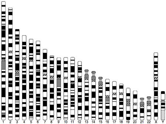
We have a total of 46 chromosomes. Half of them come from the mother and the rest, from the father. Out of these 46 chromosomes, 44 are autonomies and 2 are sex chromosomes. The sex chromosomes are not always a perfect pair.
In females there are 44 autonomies and two X chromosomes, in males there are 44 autonomies, one X chromosome and one Y chromosome. So the chromosomes
In woman are 44 + XX, while the chromosomes in man are 44 + XY. Let us see the inheritance pattern of X and Y chromosomes.
During gamete formation, the normal diploid chromosome number is halved. This is called the haploid condition. All the eggs of a female have 22 + X chromosomes. A male produces two types of sperms—one type bears the 22 + X composition and the other, 22 + Y. Therefore, in every 100 sperms, 50 have Y chromosomes and 50 have X chromosomes.
X-Y System of Sex Determination in Man
Any one of the two types of sperms can fertilize the egg. If a Y-bearing sperm fertilizes the egg, the zygote has the 44 + XY composition, and the resulting embryo grows to be a boy. When an X-bearing sperm fertilizes the egg, the resulting zygote has the 44 + XX composition. This embryo develops into a girl. All the children inherit one X chromosome from the mother.
Therefore, sex is always determined by the other sex chromosome that they inherit from the father. One who inherits the X chromosome of the father is a girl, while one who inherits the Y chromosome of the father is a boy.

Role of environment in sex determination:

Environmental conditions such as temperature around the developing embryo may determine sex in some animals. Such conditions may override the genetic basis. Some animals such as snails can even change their sex, showing that their sex is not genetically determined.

Incubation of the eggs of the turtle Chrysema picta at a high temperature produces females. But the incubation of the eggs of the lizard Agama at a high temperature produces males.
http://www.biologydiscussion.com/essay/determination-of-sex-in-human-beings/1643

Sunday 3 January 2016

B Sc II

Development of Male gametophyte (Before pollination):
Microspore or pollen grain is the first cell of the gametophyte. The microspore germinates in situ i.e. while within the microsporangium. Each microspore divides asymmetrically into a 2-cells: a smaller prothallial cell and a larger antheridialcell. The prothallial cell does not divide further while the antheridial cell divides into a smaller generative cell near the prothallial cell and a larger tube cell. Finally pollination takes place at 3-celled stage (a prothallial cell, a generative cell and a tube nucleus) (Fig. 9.12).
Cycas. A-C : Pre-pollination development of male gametophyte
Pollination:
In Cycas pollination is anemophilous (by wind). The 3-celled microspores liberate from mega-sporangia are blown away by wind. Finally microspores reach on ovules and get enlarged in the pollination drop (ooze) of micropyle. As the ooze dries up, the microspores are drawn into the pollen chamber.
(d) Development male Gametophyte (After polli­nation):
After a gap of about 4 months, post-pollination development of male gam­etophyte occurs. The exine ruptures and the intine grows out in form of apollen tube. The pollen tube acts as a haustorium, i.e. absorb food while penetrating through the nucellus and hang in the archegonial chamber. In the pollen tube, generative cell divides into a stalk cell and a body cell. Finally, the body cell divides into two male gametes or antherozoids. Thus, a fully developed male- gametophyte consists of a disorganized prothallial cell, stalk cell, tube nucleus and 2 male gametes (Fig 9.13 )
Cycas: A-F : Post Pollination development of male gametophyte
Each male gamete appears top-shaped with 5-6 spiral bands of cilia. The size of male gamete in Cycas varies from 180-210µm (largest, 400«m reported from Chigua, a cycad).
Source : http://www.biologydiscussion.com/life-cycle/life-cycle-of-cycas-vegetative-and-sexual-life-cycle/5766
Development of female gametophyte (Endosperm):
Inside the nucellus, one cell differentiated into megaspore mother cell. It undergoes reduction division (meiosis) to form a linear tetrad of four haploid megaspores. Usually, the upper 3 megaspores towards micropyle degenerate while the lower most functional megaspore (embryo sac cell) undergoes free nuclear division followed by wall formation to form a cellular female gametophyte or endosperm.
Hence, the formation of female gametophyte is monosporic, i.e develops from a single megaspore. During formation of endosperm nucellus is utilized. It should be noted that in gymnosperms the endosperm develop before fertilization and is haploid (n) while in angiosperms it is triploid (3n) and formed after fertilization (Fig. 9.10).
Development of female gametophyte of cycas within ovule
Summary of devlopment of Female Gametophyte
At the micropylar end of female gametophyte 2-8 archegonia develop. All the necks of archegonia open into an archegonial chamber formed by a depression in female gametophyte (Fig. 9.11). Each archegonium develops from single superficial cell called archegonial initial.
It gets enlarged and divides transversally into outer primary neck cell and inner central cell. The primary neck cell divides anticlinally to form two neck cells. The inner central cell enlarges and its nucleus divides into venter canal nucleus and egg nucleus. Soon the venter canal nucleus disorganizes. Thus, a mature archegonium has two neck cells and an egg. Neck canal cells are not formed. The egg cell in Cycas is largest in the plant kingdom (Fig. 9.11).
Cycas, Devlopment of archegonium
Source : http://www.biologydiscussion.com/life-cycle/life-cycle-of-cycas-vegetative-and-sexual-life-cycle/5766